Dental

Dental

Billions of people are affected by chronic caries or tooth decay. Because there is no accurate measurement that predicts if tooth decay will occur at a specific location, there are no conventional means for managing this chronic disease. For most other common chronic diseases such as atherosclerosis, high blood pressure, diabetes, and cancers, the highest risk patients are identified by measuring leading indicators of disease and managing these indicators so that surgery is held for the last resort of treatment. Currently, tooth decay does not have a leading indicator for site-specific tooth decay that can be easily measured and tracked over time.

Research: A Multimodal Intravascular Imager for Carotid Atherosclerosis

Research: A Multimodal Intravascular Imager for Carotid Atherosclerosis

Based on the unmet clinical need for improved instruments in response to the contemporary understanding of atherosclerosis biology, these researchers embarked on a quest to develop a novel platform for multimodal high-resolution structural, biochemical, and biological vascular imaging. 


Chronic Total Occlusion

Chronic Total Occlusion

Tick Tock This is a Block We Must Find a Way to Stop

Patients with Chronic Total Occlusion (CTO) of the coronary artery have lesions that create a total blockage in the heart artery—and for most, ultimately become an impediment to normal activities. Why?  Of the millions of patients, a year living with coronary artery disease, 100,000 to 200,000 patients a year are provided medication therapy--leaving their underlying lesion unaddressed.

Jim is a great example. He saw his physician because of chest pressure and shortness of breath. He was given a battery of stress tests. An angiogram revealed CTO. Because of the risk and difficulty in surgically treating the lesion, he was told medication therapy was his best option. Although he proceeded with medication, Jim, like most, continued to experience symptoms and ultimately became less active. Over time the reduced activity made him asymptomatic, so he presumed he was getting better. The reality was different. His heart muscle was working well enough to keep him alive, but he wasn’t getting better. The untreated lesion remained a ticking time bomb.

“These are the hardest lesion subsets to treat in cardiology,” says Dr. William Lombardi, an associate clinical professor of medicine in the Division of Cardiology, and the director for Complex Coronary Artery Disease Therapies in the UW Medicine Regional Heart Center. Lombardi should know. He is considered a world expert on Chronic Total Occlusion Percutaneous Coronary Intervention (CTO PCI)—and is informally referred to as “the godfather of CTO PCI”, in part because he treats patients worldwide and has done more of these procedures than anyone else in the U.S. In fact, Dr. Lombardi’s entire clinical practice was built on treating patients with coronary artery disease that have been told, “nothing more can be done”.

Dr. Lombardi explains the challenges of CTO this way: “CTO falls into a treatment paradox exacerbated by a multitude of factors, the foremost being technical complexity coupled with the lack of physician experience in percutaneously treating these lesions. This severely reduces the ranks of interventional cardiologist willing and able to provide treatment options.”

As sad as these facts are, they are somewhat understandable considering the amount of training, skill, and experience required to remove CTO lesions. Today the surgery creates anatomical ambiguity because surgeons use a two-dimensional screen to circumnavigate a three-dimensional artery. Worse, there are potentially lethal blind spots physicians encounter as they attempt to cross and treat a lesion between 1-10 cm long. Dr. Lombardi likens the experience to mining. “Imagine crawling through a mine and not being able to see where the wall or edge is—and knowing that if you touch it, the mine collapses, and causes death”.

So, what can be done to help more patients receive potentially life-saving surgical interventions that only a few physicians like Dr. Lombardi are able and willing to do?

The answer, according to Dr. Lombardi, is crystal clear: “If you can turn the lights in the artery on and provide a three-dimensional view, you can see where you are going vs. walking blindly in the dark. This reduces the guess work and visual challenges, making the procedure much less scary and more straightforward.”

And that is precisely what VerAvanti is attempting to do with the Scanning Fiber Endoscope (SFE). When asked about the potential for the SFE, Dr. Lombardi said, “It could be a real game changer because it has the potential to make the procedure safer, faster and more adoptable—in other words, instead of having a golf course for only 200 PGA tour pros, we could have a golf course that anyone in the community can play.”

Uncovering the #1 serial killer in America

Uncovering the #1 serial killer in America

Shedding light on hidden plaques causing cryptogenic stroke

Miraculously, you survived.

It was a stroke that struck without warning. Facing your mortality at just 50, sent shivers to your core. Now you have a new resolve. A vow to learn why you had a stroke, and what you can do to prevent it from happening again.

Over the next few days of your hospitalization, your physician does a workup to learn where the stroke was and to discern any patterns that could give a hint to the stroke mechanism. The search begins. It starts with a brain CT or MRI and blood tests. The heart is looked at with an EKG and Echocardiogram (ultrasound). The carotid arteries are also evaluated with US, CT or MR. 

Then your world shatters again. After all those tests, the cause of your stroke is, “undetermined”. It is called a Cryptogenic Stroke, generally due to a blockage of a brain artery by a clot. Even with the most advanced and invasive testing, including brain biopsies and prolonged cardiac monitoring, you and 250,000 others in America now share the same questions: Where did the clot come from? Will it happen again? How can it be prevented?

Uncertainty prevails, and your doctor’s explanations don't add much peace of mind. Once you’ve had this kind of stroke, the risk of recurrence is over 5.5% a year. Although very effective treatments exist for root causes of stroke, such as removing ruptured carotid plaques, the uncertainty in these cases precludes targeted therapy. You are prescribed blood thinners coupled with lifestyle changes. Given what you now know, this “shot in the dark” treatment hardly seems aggressive enough.

Cryptogenic stroke accounts for a third of all ischemic strokes (>800,000 in U.S. annually) and represents a major unresolved medical and socioeconomic problem. However, a novel diagnostic approach that could pinpoint the root cause of these strokes is just around the corner.

LUIS SAVASTANO, M.D., Clinical Advisor

LUIS SAVASTANO, M.D., Clinical Advisor

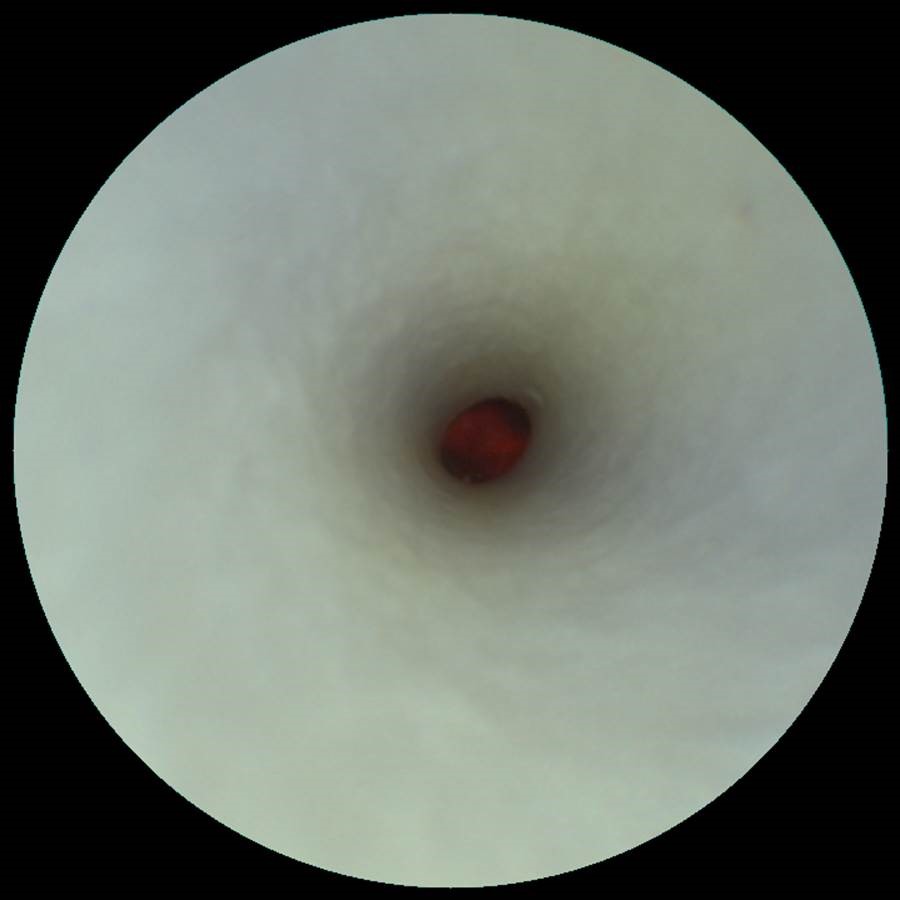
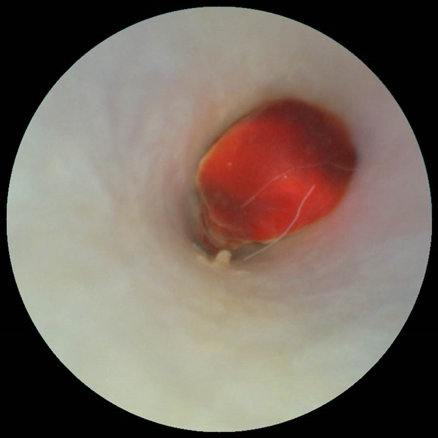
Consider the work that Luis Savastano, MD, is doing in the Department of Neurosurgery, at the University of Michigan Medicine. Dr. Savastano is a physician-scientist specialized in neurosurgery and endovascular neuroradiology. He is pioneering the use of Scanning Fiber Endoscopy (SFE) to detect hallmarks of atherosclerotic plaque rupture and vulnerability. Dr. Savastano summarizes what he has discovered this way, “The SFE can go inside the vessels and snap pictures of the carotid arteries at a very high resolution and find ‘smoking gun’ lesions, such as ruptured plaques, in patients with strokes of unknown cause. In addition, we believe the SFE may even be able to show which silent, but at-risk, plaques may cause a cardiovascular event in the future.”

His groundbreaking work, recently published in two articles in the highly respected journal Nature Biomedical Engineering, introduces SFE as a powerful multimodal platform for atherosclerosis imaging and uncovers the game changer potential for patients suspected to have ruptured but “undetectable” carotid plaques. Dr. Savastano explains, “Many times we may have a suspicion of what happens: the patient has a ruptured atherosclerotic plaque in the carotid artery (one of the most common known reasons of stroke), and it is forming clots that break and travel upstream, occluding vessels in the brain. However, seeing ‘smoking gun’ lesions in the arteries is difficult using available technologies. The majority of tools provide great pictures of the vascular lumen but have many limitations when uncovering subtle thrombogenic lesions (such as shallow ulcers and erosions) in the arterial surfaces, and consequently, many culprit plaques are wrongly labeled as innocent bystanders.” This work, supported by NIH and the Congress of Neurological Surgeons, was distinguished with the Galbraith Award for the top cerebrovascular work by the Americal Association of Neurological Surgeons.

Dr. Savastano’s team has analyzed human cadaveric and surgical tissue and found that patients suffering from cryptogenic stroke actually harbor ruptured lesions in carotid plaques that can originate clots leading to strokes. These lesions may not be detected by the imaging tools currently available, leading to misdiagnosed “cryptogenic" strokes.” These findings were presented this year at the International Stroke Conference and published in Stroke, the flagship journal of the AHA. Dr. Savastano says, “Such miss-diagnoses preclude patients from appropriate and targeted treatment, which translates into unnecessary and unrevealing tests, and a loss of opportunities to prevent future strokes.” He adds, “Angioscopy could be safely performed during workup for cryptogenic stroke, leading to the detection of the root cause of these events. Precise and timely diagnosis of a culprit carotid lesion would provide peace of mind to patients and physicians, strengthen adherence to prescribed treatments, avoid other invasive diagnostic tests (such as brain biopsies and implantable cardiac loop recorders), improve risk stratification of stroke recurrences, and provide a target for medical or surgical interventions leading to reduction of recurrence.”

If successful, Dr. Savastano believes this could ultimately change the standard of care, “Technology like the SFE will be a game-changer in the way we diagnose and treat stroke. The ability to see smoking gun lesions will enable physicians to target the management of ruptured plaques, and likely save more lives or reduce disability, all without undergoing expensive clinical workups.”

Dr. Savastano also believes that SFE provides an opportunity to potentially shift the treatment approach of atherosclerosis, the #1 killer in America, to treatments similar to breast or prostate cancer screening. He explains, “The ability of SFE to identify and monitor the biological markers that render a plaque unstable and at risk for rupture could enable the detection of individuals within high-risk populations who are most likely to suffer from cardiovascular events, and therefore benefit the most from preventive treatment during the asymptomatic stage.” He continues, “In the future, patients with significant cardiac risk factors, such as a family history of heart attacks or smokers, could have their arteries checked with the SFE, and undergo local treatment (such as stenting) if a silent but high-risk plaque is found. In addition, plaque-specific data could help physicians modulate treatment intensity of atherosclerosis, which is currently based on systemic surrogates such as cholesterol and blood sugar levels and occurrence of cardiovascular events such as stroke or myocardial infarction.”

Sample Arterial Images from Euthanized Rabbit

Dr. Luis Savastano, University of Michigan provides images obtained from a VerAvanti Research Imaging System.

Images were obtained from an euthanized rabbit.

Looking at the inside of the catheter.

Looking at the inside of the catheter.

Inside the artery (away from thrombus)

Inside the artery (away from thrombus)

Inside the artery (close up of thrombus)

Inside the artery (close up of thrombus)

Bifurcation after removing the thrombus.

Bifurcation after removing the thrombus.

Sample Arterial Images from Post Mortem Human

Dr. Luis Savastano, University of Michigan provides images obtained from a VerAvanti Research Imaging System.

Images were obtained from post mortem human arteries.

Healthy Endovascular Surface

Healthy Endovascular Surface

Uncomplicated Artheroma at the Carotid Bifurcation

Uncomplicated Artheroma at the Carotid Bifurcation

Nature, Biomedical Engineering (volume 1, article 0023)

Nature, Biomedical Engineering (volume 1, article 0023)

Multimodal laser-based angioscopy for structural, chemical and biological imaging of atherosclerosis

Multimodal SFE video acquisition of post-mortem arteries. Endoluminal endoscopic videos (Supplementary Videos 1,2,3,4,5) were obtained by navigating the SFE probe into the common carotid artery (CCA) toward the CB and then into the first 2 cm of the ICA and external carotid artery (ECA).

Increased Accuracy of Atherosclerosis Detection Using Scanning Fiber Endoscope

Increased Accuracy of Atherosclerosis Detection Using Scanning Fiber Endoscope

Researchers from the University of Michigan used a unique application of a medical camera to view the carotid artery to assess the risk of atherosclerosis. According to researchers at the University of Michigan School of Medicine used a scanning fiber endoscope, or SFE, to acquire high-quality images of potential atherosclerosis regions of the carotid artery that can be missed by conventional radiological techniques. "The camera actually goes inside the vessels," Dr. Luis Savastano is a Michigan Medicine resident neurosurgeon and first author of the study. He said in a press release, "We can see with very high resolution the surface of the vessels and any lesions, such as a ruptured plaque, that could cause a stroke. This technology may even be able to show which silent, but at-risk, plaques may cause a cardiovascular event in the future."